Research News

T. Rex Had Osteomyelitis!

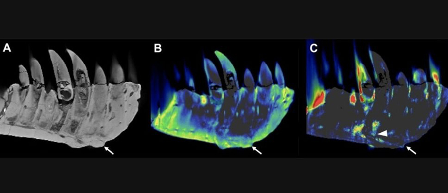
Radiologists in Germany used dual-energy CT to diagnose osteomyelitis in a famous dinosaur

(A) Conventional CT images show a lateral view of the tooth-bearing part of the left dentary. The arrow indicates an abnormal growth that sticks out from the surface of the tissue. (B) The DECT-based calcium material map shows a homogeneous mineral distribution, while (C) the fluorine material map shows significant mineral accumulation in the center of the abnormal growth and adjacent tooth roots (arrowhead). (Image credit: RSNA and Charlie Hamm, M.D.)

Dual-energy CT (DECT) was recently used by radiologists to diagnose a 63 million year-old Tyrannosaurus rex dinosaur, according to a recent abstract at the Radiological Society of North America. Dr. Charlie Hamm, a radiologist at the Charité University Hospital in Berlin, and his team used DECT to scan the dinosaur’s mandible, measuring 31.3 inches (79.5 centimeters) long and had a maximum thickness of 3.2 inches (8.1 cm).

Check out the abstract and a LiveScience article on the research

Happy New Year to all members and friends of the ACVR!


Photograph of the fossilized T. rex named Tristian Otto, diagnosed with mandibular osteomyelitis. (Image credit: RSNA and Charlie Hamm, M.D.)