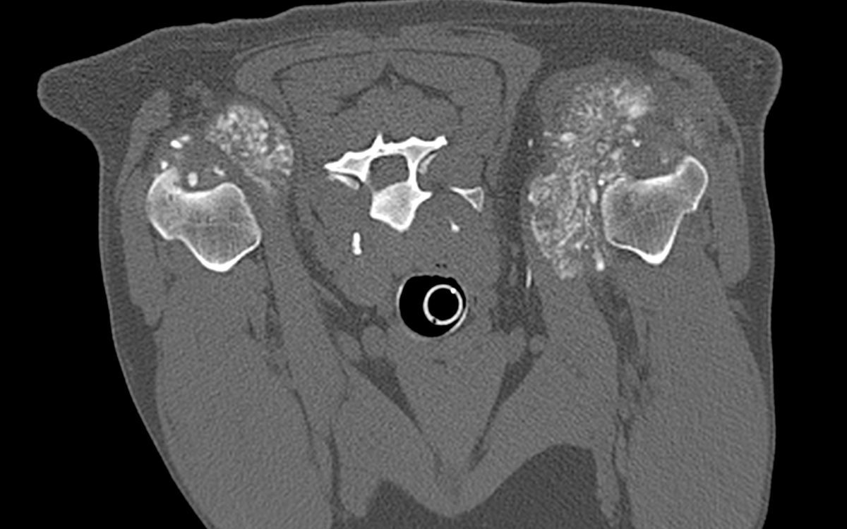
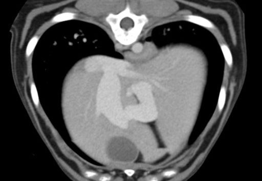
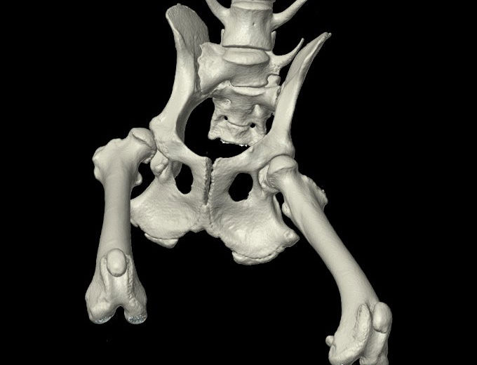
Computed Tomography (CT) imaging uses X-rays in conjunction with computing algorithms to image the body. In CT, an X-ray generating tube opposite an X-ray detector (or detectors) in a ring shaped apparatus rotate around a patient producing a computer generated cross-sectional image (tomogram). Radiocontrast agents are often used with CT for enhanced delineation of anatomy and angiography. With computer manipulation, CT images can be reconstructed into 3D images. Faster scanning times in modern equipment has been associated with increased utilization. (Adapted from Wikipedia. Read More…)
Because CT can produce harmful ionizing radiation, the ACVR supports sustained and conscientious safety practices as detailed in our Radiation Safety Statement.