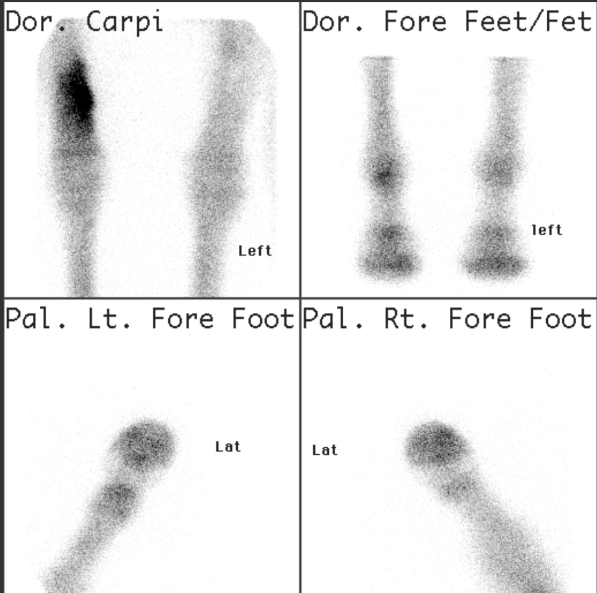
Bone scintigraphy in a horse with a radial stress fracture
Nuclear medicine imaging involves the administration into the patient of radiopharmaceuticals consisting of substances with affinity for certain body tissues labeled with radioactive tracer. The most commonly used tracers are Technetium-99m, Iodine-123, Iodine-131 and Xenon-133. The heart, lungs, thyroid, liver, gallbladder, and bones are commonly evaluated for particular conditions using these techniques. While anatomical detail is limited in these studies, nuclear medicine is useful in displaying physiological function. The excretory function of the kidneys, iodine concentrating ability of the thyroid, blood flow to heart muscle, etc. can be measured. (Adapted from Wikipedia. Read More…)
Because nuclear medicine imaging can produce harmful ionizing radiation, the ACVR supports sustained and conscientious safety practices as detailed in our Radiation Safety Statement.