-
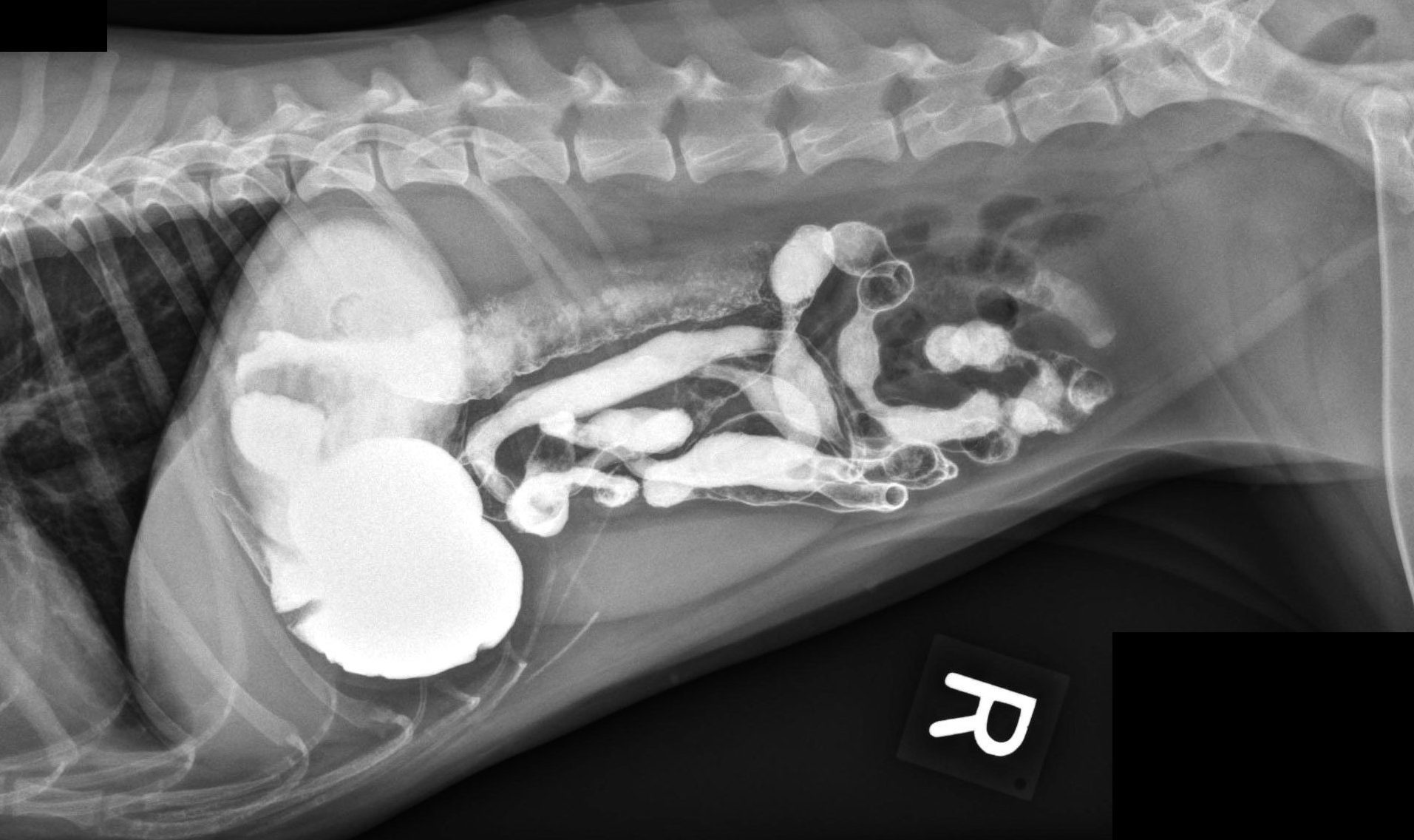 Abdominal radiograph of a dog after administration of barium (GI series)
 -
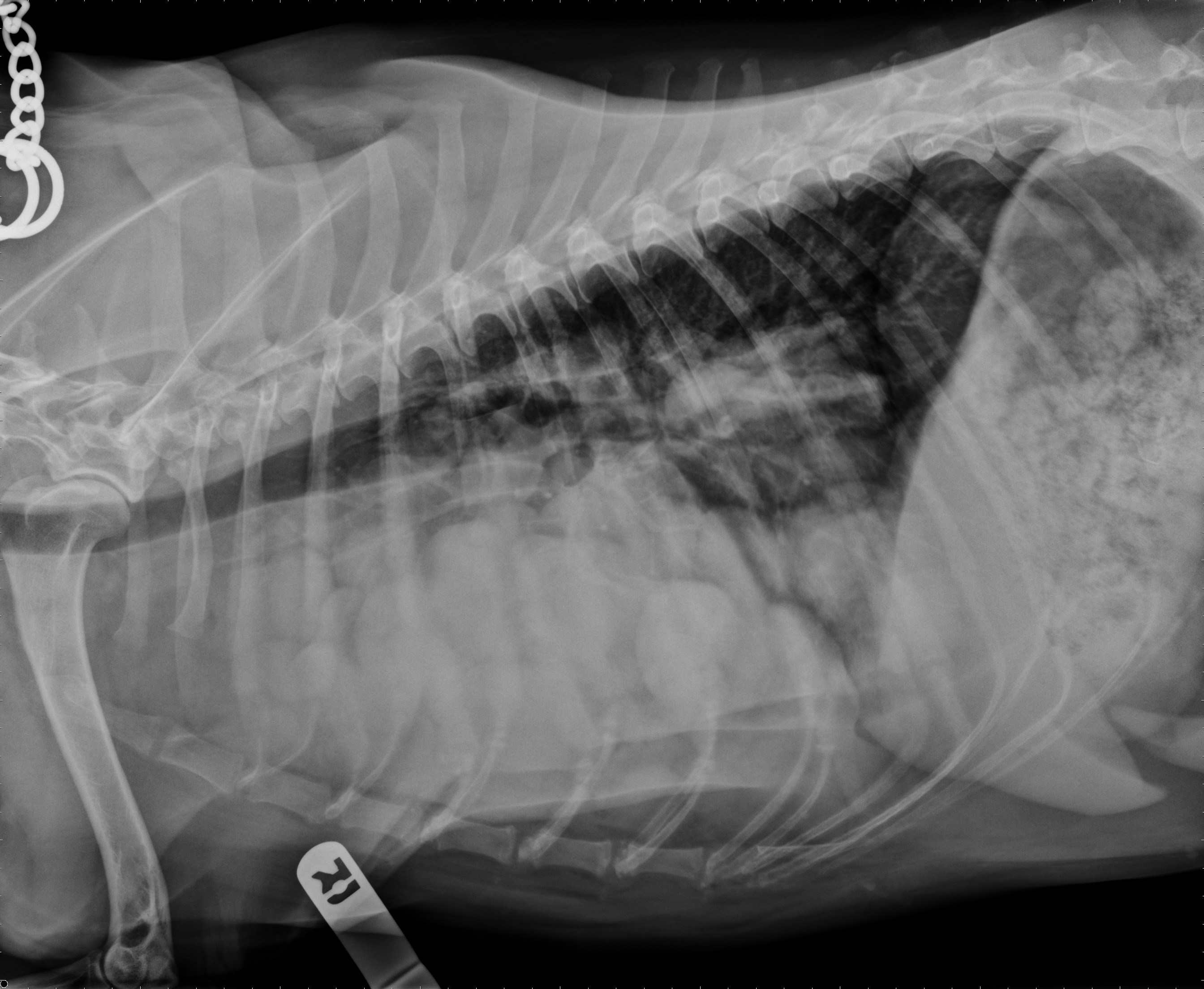 Thoracic radiograph of a dog with mediastinal masses (thymoma)
 -
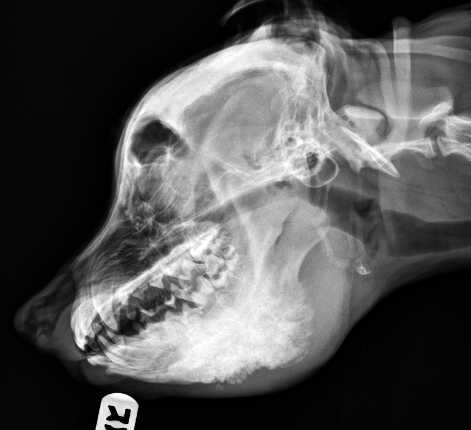 Lateral skull radiography. Diagnosis: Craniomandibular Osteopathy (CMO)
 -
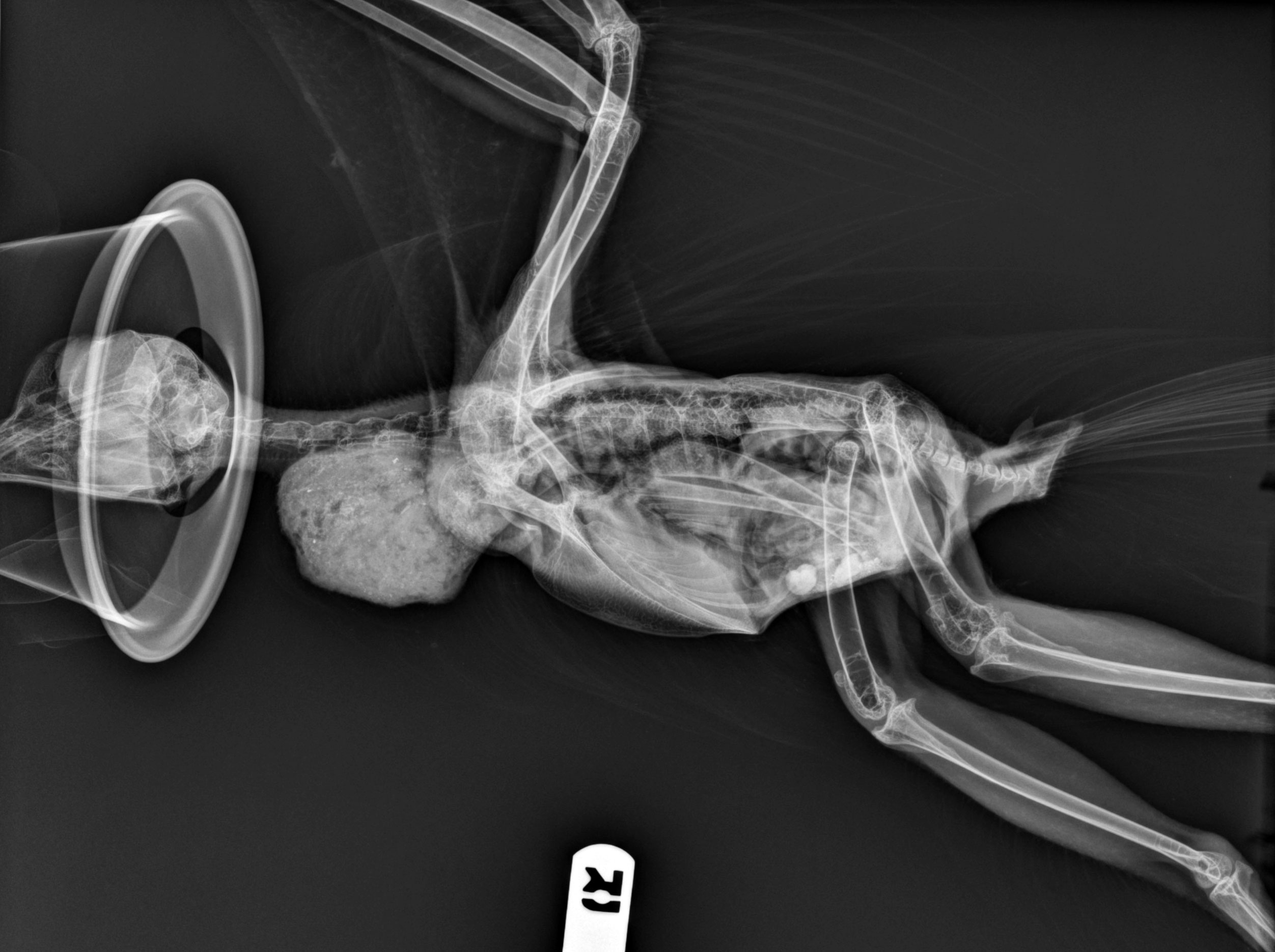 Lateral radiograph of a hawk after ingestion of large fragments of bone
 -
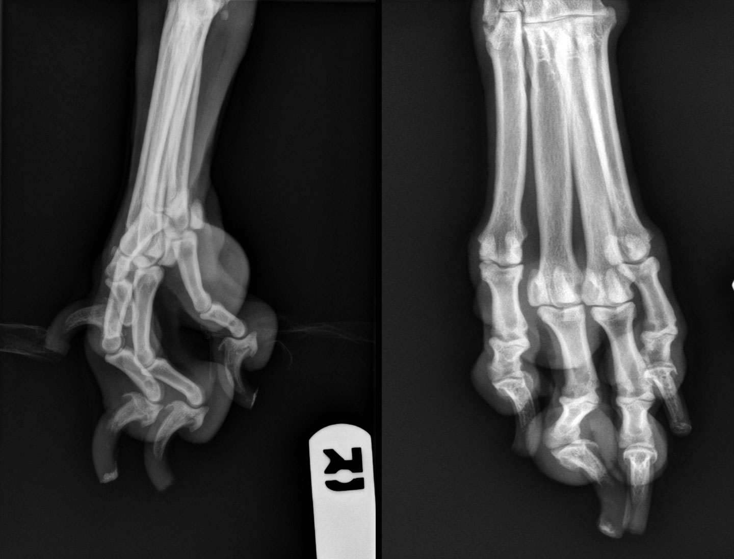 Radiographs of a dog's paw. Diagnosis: Digital tumor (subungual carcinoma)
Radiology is the specialty of directing medical imaging technologies to diagnose and sometimes treat diseases. Radiography involves the use of x-rays to produce radiographs. Radiography is the oldest but most common modality used in medical imaging.
After extensive training, radiologists also direct other imaging technologies such as ultrasound, computed tomography (CT), magnetic resonance imaging (MRI) and Nuclear Medicine to diagnose or treat disease. (Adapted from Wikipedia. Read More..)
Because radiographs can produce harmful ionizing radiation, the ACVR supports sustained and conscientious safety practices as detailed in our Radiation Safety Statement.
ACVR’s Radiation Safety Statement
The ACVR supports sustained and conscientious attention to safe practices regarding veterinary radiologic imaging and therapy as they relate to personnel, patient, and equipment. Exposure to radiation should always be As Low As Reasonably Achievable (ALARA) while maximizing the quality of the procedure.
The use of radiation to diagnose and treat animal diseases has significantly advanced the field of veterinary medicine, resulting in improved patient care. The equipment and techniques used to perform imaging and therapy procedures, whether they generate ionizing radiation (such as radiography, fluoroscopy, computed tomography, therapeutic radiation generators, and radioisotopes) or nonionizing radiation (such as ultrasonography and magnetic resonance imaging) are associated with potential risks to patients and personnel. Individuals with significantly less formal training than ACVR Diplomates in the safety, physics, and biology of ionizing radiation are routinely involved with these procedures. It is essential that these individuals be adequately trained in the appropriate function and use of the equipment and in the techniques of the procedure to minimize unnecessary radiation exposure to themselves, staff, patients, and the public.
Facilities employing ionizing radiation* as a diagnostic and therapeutic tool must have established policies and procedures in place and be familiar with current standards and techniques. This includes developing examination protocols for radiography, fluoroscopy, computed tomography, and radioisotope procedures that take into account patient body parameters (size, weight, composition) and maximize the distance and shielding of workers and the public. Equipment quality control must be included in these protocols.
Veterinary medical personnel who are directly involved with ionizing radiation procedures should wear dosimetry badges to monitor radiation exposure. Badge measurements should be evaluated regularly. If individual doses are reported to be high, measures should be taken to reduce further exposure to that individual (e.g., minimizing exposure time, maximizing distance, wearing appropriate shielding). A human body part within a primary x-ray beam is a failure in safety protocol and should be prevented through proper training and chemical restraint of the patient when necessary.
*Ionizing radiation refers to any electromagnetic or particulate radiation capable of producing ion pairs by interaction with matter. Common examples used in medicine include x-rays, electrons, gamma rays, beta particles, and other products of radioactive decay.
Seleced References:
- U.S. Nuclear Regulatory Commission – Occupational dose limits for adults.
- National Council on Radiation Protection and Measurements (NCRP) – NCRP Report No. 148, “Radiation Protection in Veterinary Medicine” (ISBN 0-929600-85-1) was published in 2004. This report can be purchased ($50 hardcopy; $40 PDF) from the NCRP web site.
- Health Canada – Environmental and Workplace Health – Radiation Protection In Veterinary Medicine – Recommended Safety Procedures For Installation And Use Of Veterinary X-ray Equipment – Safety Code28.
- Health Canada – Environmental and Workplace Health – X-rays.
Lower the Dose Initiative

The ACVR partners with IDEXX and the National Association of Veterinary Technicians in America (NAVTA) in Lower the Dose, a new initiative to help raise awareness of radiation safety best practices. There is a need for more radiation safety education to make people aware of the As Low as Reasonably Achievable (ALARA) principle to help create a safer work space. Visit the new Lower the Dose website for timely and relevant radiation resources, news, safety information, and state guidelines. This initiative complements the ACVR’s Position Statement on Radiation Safety.
Image Resolution & DICOM Guidelines
The following recommendations formulated by the ACVR Digital Imaging Standards Committee (DISC) have been adopted by the ACVR as guidelines for the veterinary profession:
1. The ACVR considers 2.5 lp/mm as the mininum standard for spatial resolution in a clinical setting for primary capture digital radiographic devices. This recommendation also applies to secondary capture of radiographic images such as digital cameras, laser scanners, etc.
2. The ACVR recommends defining a DICOM study as follows:
Computed Tomography/Magnetic Resonance Imaging (CT/MRI)
- Localizer images (scout/topograms) should constitute a single series and should not be part of an axial dataset.
- A group of axial images acquired in a contiguous fashion should constitute a series.
- Reconstructed datasets should comprise a separate series.
Ultrasound (US)
- Still images should all be in the same series. Each cineloop should comprise one series.
Radiography (CR, DX, OT, SC)
- A study should comprise a group of images of the same region taken at radiographic examination (e.g., a 3-view thoracic examination constitutes a DICOM study).
- Each image taken in the radiographic examination should constitute a separate series.
- Images modified in post processing should be in the same series as the original image.
Adding images to a closed study (i.e., a study already sent to the archive or telemedicine service for review) is discouraged. While the addition of images to a prior study is often convenient for the clinician, this can potentially result in submission of images to a consultant after a report has been generated on that study. The additional images may contain radiographic findings that may compromise the accuracy of the report. Therefore adding images to a closed study is discouraged.